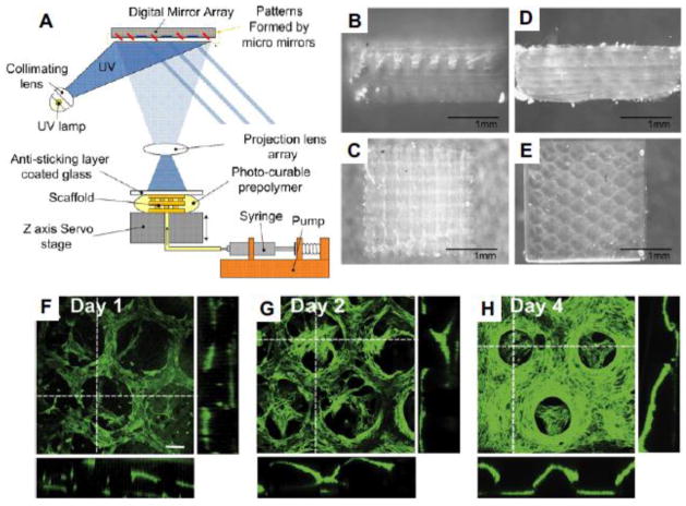
Figure 2. Stereolithographic bioprinting.
(A) Schematic illustration of stereolithography system (Gauvin et al. 2012). (B)–(E) The side view of woodpile (B) and hexagonal (D), as well as the top view of woodpile (C) and hexagonal (E) structures generated by their stereolithography system. (F)–(H) 3D confocal images showing the proliferation of encapsulated HUVEC cells in day 1 (F), day 2 (G) and day 4 (H). (Scale bar: 100 μm)

