
Figure 1.
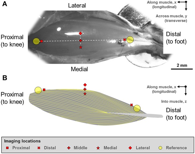
(A) Digital photograph of the mouse tibialis anterior (TA) muscle. (B) Schematic illustration of the muscle architecture in the mid-sagittal plane of the muscle (Heemskerk et al., 2005; Lovering et al., 2013). The fiber orientation is indicated by the yellow outlines. The sarcomere lengths of the mouse tibialis anterior muscle were imaged at five defined sites. Two reference points were first defined at the proximal and distal ends of the TA muscle. From these reference points, five imaging sites located along the longitudinal axis (“proximal,” “middle,” “distal” sites) and along the transverse axis (“medial,” “middle,” “lateral” sites) were identified on TA. The “middle” landmark was the mid-point of the straight line connecting the “proximal” and “distal” TA sites, while the “medial” and “lateral” landmarks were ~500 μm away from the “middle” landmark in the medial and lateral directions, respectively.