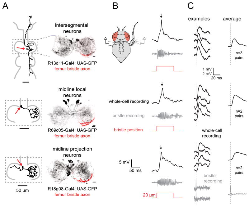
Figure 3. Three classes of VNC neurons that receive direct synaptic input from the same femur bristle.
(A) Left: morphology of a biocytin-filled neuron which expressed GFP in the indicated genotype. Red arrows indicate cell body position. Right: maximum intensity projection of GFP expression within the prothoracic neuromere of the VNC (black), co-labeled with the axonal arbor of a single femur bristle neuron filled with DiI (red); we always targeted the same femur bristle (Figure S5A). All three central neuron classes overlap with this bristle neuron axon. Scale bars, 10 Im. See also Figures S3–S5.
(B) Each row shows a typical in vivo whole-cell current-clamp recording from a central neuron and the simultaneously-recorded signal from a bristle neuron. As before, we targeted the same bristle on the femur, near the femur-tibia joint (Figure S5A).
(C) Single spikes in this bristle neuron reliably trigger excitatory postsynaptic potentials (EPSPs) in each class of central neuron. As before, we targeted the same bristle on the posterior femur, near the femur-tibia joint (Figure S5A). Examples of bristle neuron spikes are shown at bottom. The left column shows representative single-trial EPSPs recorded from each corresponding central neuron class. At right are spike-triggered-averages of the postsynaptic voltage, averaged across all paired recordings from the same central neuron class where a connection was detected.

