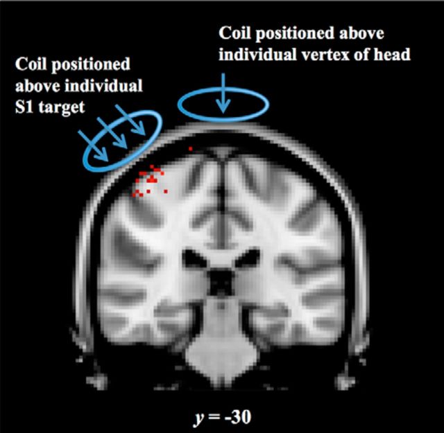
Figure 2.
rTMS target placement. Each point represents an individual subject's right hemisphere primary somatosensory cortex S1 target (red) transformed into MNI space. The S1 target was individually selected based on the peak of the subject's Z-statistic map of brushing > rest. The coil was aligned in the posterior-anterior direction at ∼45° to the right of midline in the S1 condition. In the vertex condition, the coil was placed directly above the interhemispheric fissure in the same coronal plane as individual subject's S1 target. The coil was aligned in the posterior-anterior direction at 0° to the midline. Each target is displayed at the section representing the group average y-coordinate (coronal) in MNI space.

