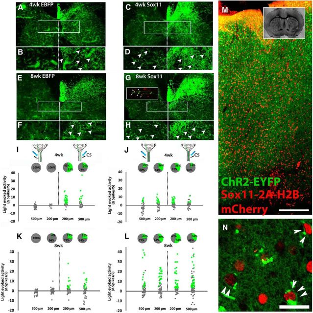
Figure 4.
Sox11-transduced CST axons sprout across the spinal midline after pyramidotomy and form functional synapses with spinal neurons. Animals received unilateral pyramidotomy and cortical injection of mixed EBFP/ChR2–EYFP or Sox11/ChR2–EYFP to the uninjured cortex. Four or 8 weeks later, Sox11-treated (C, D, G, H) but not EBFP-treated animals (A, B, E, F) showed robust midline crossing of CST axons in the cervical spinal cord. Crossed CST axons colocalized with VGluT1 (red, arrowheads, inset in G). I, J, Optical stimulation of the right and left spinal cord was paired with single-unit extracellular recording. In EBFP control animals, activity was significantly increased by optical stimulation in units located on the right, but not left, spinal cord (green dots, I and K). Pie charts show the percentage of cells that significantly increased firing rate during light stimulation. Sox11 animals showed significant elevation of firing rates in both right and left spinal cord, indicating activation of spinal units by selective optical stimulation of CST axons that sprouted across the midline. Green indicates >2 spikes/s increase, p values <0.0001, paired t test, n = 4 animals per treatment. M, N, Confocal images of cortex 8 weeks after transduction with ChR2–EYFP (green) and Sox11–2A–H2B–mCherry (red). ChR2–EYFP was diffusely expressed but at high magnification was detectable in cellular membranes (arrowheads, N), 86.2% of which coexpressed nuclear localized mCherry. More than 300 cells from three animals were quantified. Scale bars: M, 250 μm; N, 20 μm.

