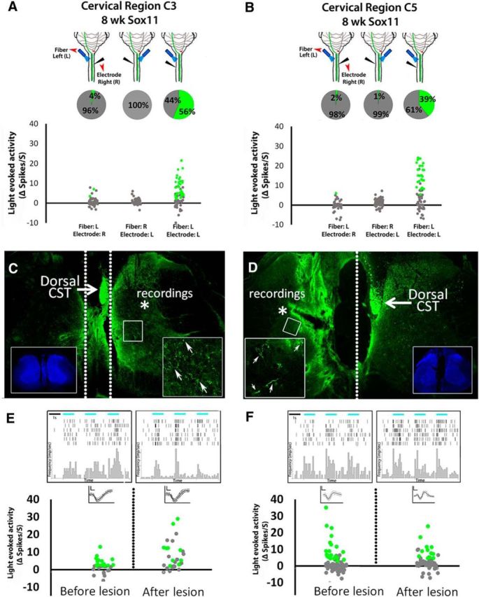
Figure 5.

Spinal activity evoked by optical stimulation of cross-midline CST terminals arises from local synaptic connections and not scattered light or antidromic stimulation of commissural relay circuits. A, B, In Sox11-treated animals with confirmed crossing of CST axons from right to left spinal cord; recording electrodes and optical fibers delivering 473 nm light were variably positioned in the right or left spinal cord at C3 (A) or C5 (B). Pie charts show the percentage of cells that significantly increased firing rate during light stimulation. Optical stimulation evoked recorded activity only when both the electrode and the light stimulus were located ipsilaterally, demonstrating local as opposed to relayed spinal responses (dotted lines, C, D). C–F, Animals received cortical injections of AAV–ChR2–EYFP and AAV–EBFP or AAV–Sox11. Eight weeks later, optical stimulation of the cervical spinal was confirmed to evoke significant postsynaptic activity, and then complete sagittal transections were acutely performed to separate the main CST from recording sites (dotted lines, C, D). Lesion completeness was confirmed in transverse sections (C, D, insets show nuclear stain). After lesion, optical stimulation of terminals continued to evoke significant increases in firing rates (peri-event raster histogram, E and F; green indicates p < 0.0001, paired t test), confirming the ability of isolated CST terminals to modulate postsynaptic firing in the absence of input from contralateral spinal cord.
