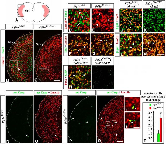
Figure 11.
Lack of Pax2+ GABAergic neurons in the SpV nuclei of E18.5 Ptf1a−/− embryos. Transverse sections of caudal hindbrain with genotypes and antibody markers indicated. Images in B, C, and N–Q correspond to the area boxed in the schematic A. High-magnification images correspond to regions boxed in adjacent data panels. B–E, Both Lmx1b+ (B, D, arrowheads) and Pax2+ (B, D, arrows) SpV neurons were detected in controls, but only Lmx1b+ SpV neurons (C, E, arrowheads) were present in Ptf1a−/− mutants. F, G, Lmx1b+ SpV neurons coexpressed Tlx3 in both controls and Ptf1a−/− mutants (arrowheads). H, I, In the SpV nuclei of Gad67-GFP mice, Pax2+ cells coexpressed GFP (H, arrowheads), suggesting that they were GABAergic neurons. I, In Ptf1a−/− Gad67-GFP mutants, GFP signal was absent. J–M, In the Ptf1aCre/+;nLacZ (control) SpV nuclei, some β-gal+ cells coexpress Pax2 (J, arrowheads), while others coexpress Lmx1b (L, arrowheads). In Ptf1aCre/YFP;nLacZ (Ptf1a−/−) mutants, β-gal+ cells do not express Pax2 (K), but the vast majority of them express Lmx1b (M, arrowheads). N–T, More Casp3+ apoptotic cells (arrowheads) were present in the SpV region of Ptf1a−/− mutants relative to controls (T, p = 0.003). In Ptf1a−/− mutants, some apoptotic cells coexpress Lmx1b (P, Q, r, s, arrowheads). Scale bars: B, C, N–Q, 100 μm; D–M, 40 μm; r, s, 20 μm.

