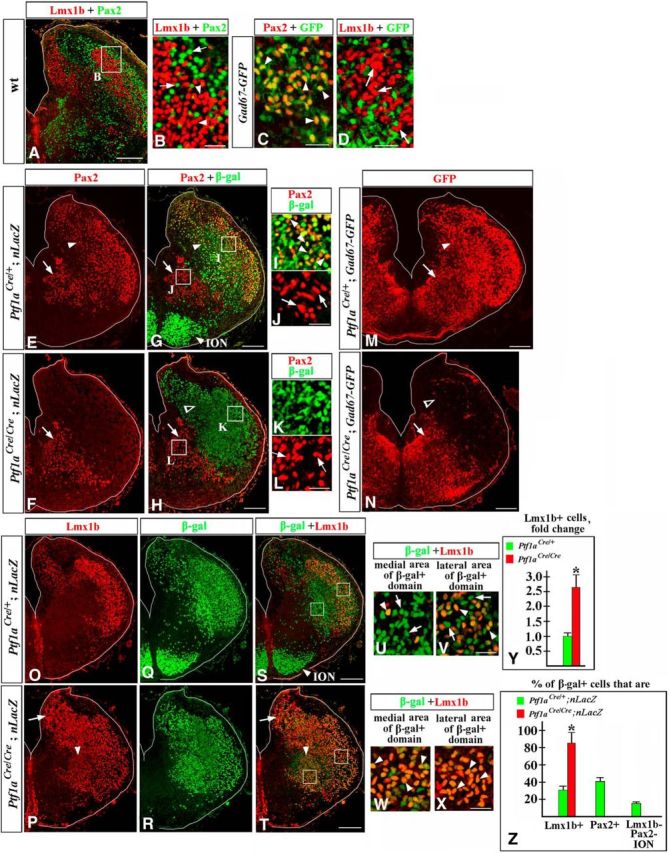
Figure 7.

Misspecified neurons in E14.5 Ptf1a−/− rh7. Transverse sections of E14.5 rh7 with genotypes and antibody markers indicated. A–D, Lmx1b and Pax2 are expressed in different sets of cells. B–D show higher magnification of cells located in dorsolateral rh7 (e.g., area indicated by a box in a lower-magnification panel, A). B, Arrowheads point to Lmx1b+ cells, arrows point to Pax2+ cells. C, D, In Gad67-GFP rh7, Pax2+ cells express GFP (C, arrowheads), while Lmx1b+ cells do not (D, arrows), suggesting that Pax2+, but not Lmx1b+, cells are GABAergic. E–L, Ptf1aCre/+;nLacZ (E, G, I, J) and Ptf1aCre/Cre;nLacZ (F, H, K, L) rh7 immunostained against Pax2 and β-gal. Magnified regions (I–L) correspond to areas boxed in G and H. E, G, I, J, In wild-type rh7, Pax2+ dorsolateral cells are β-gal+ (E, G, I, arrowheads) and thus, originate from Ptf1a-expressing progenitors. Pax2+ ventromedial cells are β-gal- (E, G, J, arrows) and originate from progenitors that do not express Ptf1a. F, H, K, L, In Ptf1aCre/Cre;nLacZ (Ptf1a−/−) embryos, β-gal+ cells, populating dorsolateral rh7, do not express Pax2 (F, H, open arrowhead, K), while the ventromedial group of Pax2+ cells is not affected (F, H, L, arrows). M, N, In Ptf1a−/− Gad67-GFP mutants, GFP staining is lost in dorsolateral (N, open arrowhead) but is preserved in ventromedial (arrow) rh7. O–X, Low (O–T) and high (U–X) magnification of Ptf1aCre/+;nLacZ (O, Q, S, U, V) and Ptf1aCre/Cre;nLacZ (P, R, T, W, X) rh7 immunostained against Lmx1b and β-gal. U and W show cells located in ventromedial rh7 (such as area indicated by left box in S and T). V and X show cells located in dorsolateral rh7 (such as area indicated by right box in S and T). O–Z, In Ptf1aCre/+;nLacZ control embryos, a fraction of β-gal+ cells (arrowheads) coexpress Lmx1b (O, Q, S, yellow cells). Approximately a half of β-gal+ cells were Lmx1b+ in lateral rh7 (V, arrowheads), while in more medial rh7, virtually all β-gal+ cells were Lmx1b− (U, arrows). In Ptf1aCre/Cre;nLacZ (Ptf1a−/−) embryos, a vast majority of β-gal+ cells coexpress Lmx1b (P, R, T), including those populating both medial and lateral rh7 (W, X, arrowheads). Y, Cell counts revealed a dramatic increase in the number of Lmx1b+ cells in E14.5 Ptf1a−/− rh7 compared with control littermates (p = 0.006). Excessive Lmx1b+ cells were particularly obvious in dorsomedial (P, T, arrow) and ventromedial (P, T, arrowhead) areas of rh7. Z, Quantification of the fractions of β-gal+ cells expressing specific markers in Ptf1a−/− and control littermates. Approximately 85% of β-gal+ cells coexpressed Lmx1b in Ptf1aCre/Cre/;nLacZ (Ptf1a−/−) rh7 vs 32% of β-gal+ cells in Ptf1aCre/+;nLacZ (control) rh7 (p = 0.001). In Ptf1aCre/+;nLacZ (control) rh7, 41% of β-gal+ cells expressed Pax2, and ∼16% of β-gal+ cells were located in the ION and did not express either Pax2 or Lmx1b. In Ptf1aCre/Cre//nLacZ (Ptf1a−/−) mutants, β-gal+ cells were not present in the ION, and none of the β-gal+ cells expressed Pax2. Scale bars: A, E, G, F, H, M–T, 200 μm; B–D, I–L, U–X, 30 μm.
