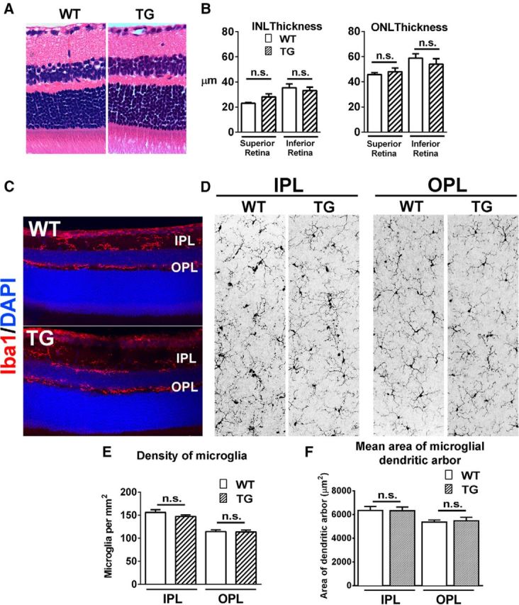
Figure 1.

Adult TG mice demonstrate normal retinal thickness and lamination and normal microglial morphology and distribution. H&E-stained paraffin sections of 2-month-old adult C57BL/6J WT and TG mice, demonstrate similar retinal structure and lamination (A), and have no significant differences in inner nuclear layer (INL) and outer nuclear layer (ONL) thicknesses (B). Microglia in the retina of animals of both genotypes demonstrated a similar laminar distribution of Iba1+ (red) microglia in the IPL and OPL (C). En face inspection of Iba1+ retinal microglia in flat-mounted retina demonstrates similar ramified morphologies in the IPL and OPL layers (D), with similar mean densities in the plexiform layers (E) and similar mean size of individual microglial dendritic arbors. Graphical data are represented as mean ± SEM; data from three female animals in each group in B and E, from four female animals in F. (n.s. indicates comparisons for which p > 0.05, unpaired t test with Welch's correction).
