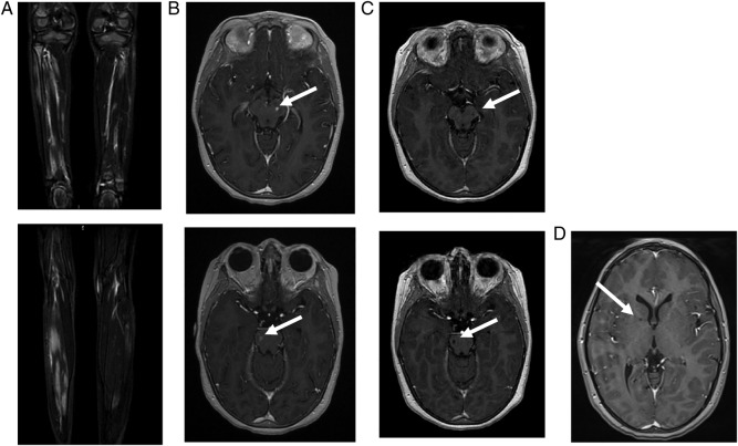
Figure 1.
Imaging of patient 1. (A) Muscle MRI of both legs performed at the age of 5 years: coronal gadolinium-enhanced T1-weighted sequence at different levels, showing patchy inflammatory muscular lesions. (B) Cerebral MRI performed at the age of 7 years. Left: axial gadolinium-enhanced T1-weighted sequence, showing gadolinium-enhanced mesencephalic (up) and peduncular (down) lesions. Right: axial diffusion-weighted sequence showing mesencephalic (up) and peduncular (down) hyperintensities. (C) Cerebral MRI performed 4 months later: axial gadolinium-enhanced T1-weighted sequence showing evolution towards lacunar lesions (up: mesencephalic lesions/down: peduncular lesions). (D) Cerebral MRI performed 3 years later: gadolinium-enhanced T1-weighted sequences showing lacunar lesions located in the internal capsule.

