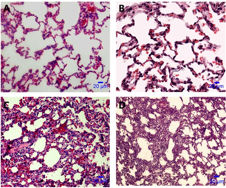
FIG 4.
Microscopic appearance of an A. baumannii-instilled lung by light microscopy (hematoxylin-eosin stain; magnification, ×400). (A) Normal microscopic appearance of lungs after saline instillation. (B) C. albicans airway colonization; lungs appear similar to those in the control group. (C) A. baumannii infection in the absence of prior Candida colonization; inflammatory-cell infiltration and alveolar damage. (D) C. albicans plus A. baumannii infection; heavier infiltration of inflammatory cells and alveolar damage than with A. baumannii infection in the absence of prior C. albicans colonization (panel C).

