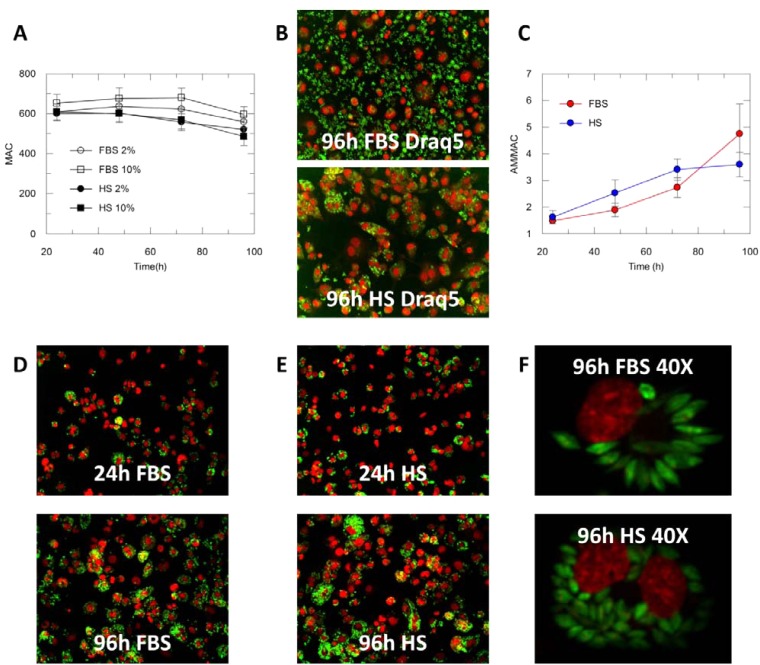
FIG 1.
(A) Average number of THP-1 cells (6 fields) in assay media containing 2% FBS, 10% FBS, 2% HS, 10% FBS. (B) THP-1 cells (Draq5, red) infected with L. donovani (green) in the presence of FBS 2% or HS 2% at 96 h (20×, air objective. (C) Evolution of the number of amastigotes per macrophage (AM/MAC) in the presence of 2% HS (blue) or 2% FBS (red) and 0.5% DMSO. Number of amastigotes per total macrophages is represented; final percentage of infection: 86% in the presence of FBS and 78% in the presence of HS. (D) THP-1 cells (DAPI, red) infected with L. donovani (green) in the presence of 2% FBS at 24 and 96 h (20×, air objective). (E) THP-1 cells (DAPI, red) infected with L. donovani (green) in the presence of 2% HS at 24 and 96 h (20×, air objective). (F) THP-1 cells (DAPI, red) infected with L. donovani (green) in the presence of 2% FBS or 2% HS at 96 h (40×, water objective).

