Figure 3.

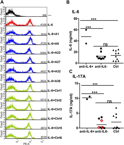
Testing for biological effects of IL‐6 antibodies (A) intracellular phospho‐STAT3 staining in cells incubated with medium alone (black), or plus IL‐6 (red) or plus IL‐6 pre‐incubated with either APECED patients’ IgG (blue) or controls’ IgG (green). Serum concentrations of IL‐6 (B) and IL‐17A (C) in APECED patients with or without IL‐6‐specific autoantibodies and in age‐matched healthy controls. APECED patients positive for anti‐IL‐17A autoantibodies are indicated with red open diamonds.
