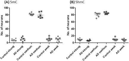
Figure 2.

Semi‐quantitative analysis of global 5‐methylcytosine (A, 5mC) and 5‐hydroxymethylcytosine (B, 5hmC) immunohistochemistry in AD and normal controls. No significant difference were detected in numbers of strong, medium and weakly stained neuronal nuclei counted in the entorhinal cortex from AD vs. normal control cases.
