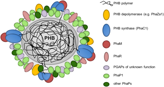
Figure 8. Model of an in vivo PHB granule in R. eutropha H16.
The surface layer is free of phospholipids and consists of proteins only. The presently known PHB granule associated proteins (PGAPs) are symbolised by differentially coloured globules. All proteins in this model had been previously shown to be bound to PHB granules in vivo by expression of appropriate fusions with fluorescent proteins. For details and overview see references8,26. The dimension of the surface layer is enlarged relative to the polymer core for better visibility.

