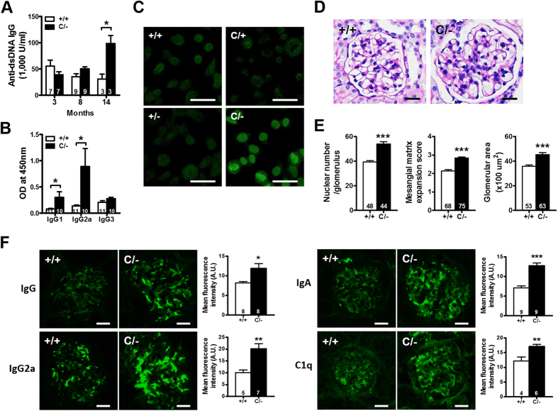
Figure 3. The development of autoimmune disorders in older PPARγ hypomorphic mice.
(A) Antibodies against dsDNA in the sera from mice at different ages. (B) IgG subclass of anti-dsDNA in the sera of 14-mo-old mice. (C) HEp-2 cells stained for anti-nuclear antibodies from the sera of 14-mo-old mice. Scale bar, 40 μm. (D) Representative periodic acid-Schiff (PAS)-stained glomerular morphology and (E) histomorphometric quantification of 14-mo-old mice. Scale bar, 20 μm. (F) Representative immunofluorescent images and quantifications of immune complex deposition for IgG, IgG2a, IgA and C1q in the glomeruli of 14-mo-old mice. Scale bar, 20 μm. Relative mean fluorescence intensity was analyzed using ImageJ software. Numbers inside bars indicate the number for each group. *p < 0.05; **p < 0.01; ***p < 0.001.

