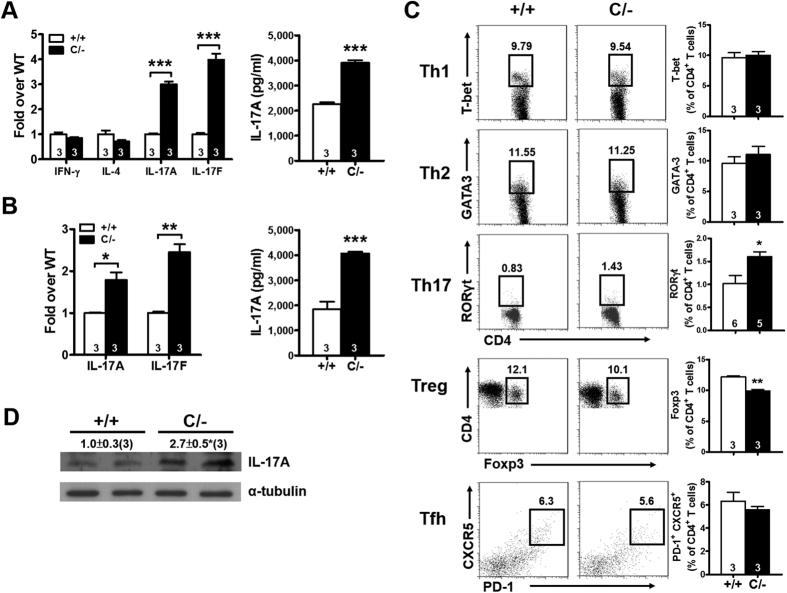
Figure 5. Increased Th17 population in the spleen of PPARγ hypomorphic mice.
(A) Relative expression of cytokines in the splenic CD4+ T cells and the protein levels of IL-17A in the medium stimulated with anti-CD3/CD28 for 3 d. (B) Relative expression of IL-17 in the splenic CD4+ T cells and the protein levels of IL-17A in the medium stimulated with TGF-β (5 ng/ml) and IL-6 (20 ng/ml) for 3 d. (C) Flow cytometric analysis of transcription factors (T-bet, GATA-3, RORγt, and Foxp3) in the splenic CD4+ T cells and expression of PD-1 and CXCR5 on the splenic CD4+ T cells. (D) Immunoblotting of IL-17A in the spleen. Mice are 4–6-mo-old. Numbers inside bars or parentheses indicate the number for each group. *p < 0.05; **p < 0.01; ***p < 0.001.

