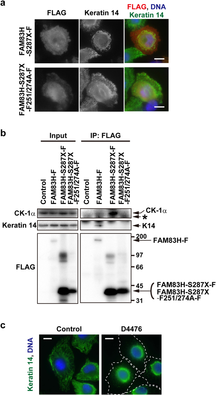
Figure 4. A truncated mutant of FAM83H disorganizes the keratin cytoskeleton in a manner dependent on its binding to CK-1 in HAM2 cells.
(a) HAM2 cells were transfected with FAM83H-S287X-FLAG or FAM83H-S287X-F251/274A-FLAG and co-stained with anti-FLAG and anti-keratin 14 antibodies. (b) A co-immunoprecipitation assay with an anti-FLAG antibody was performed using lysates from HAM2 cells transfected with FAM83H-FLAG, FAM83H-S287X-FLAG, FAM83H-S287X-F251/274A-FLAG, or the empty vector (control). Input lysates and immunoprecipitates (IP) were analyzed by Western blotting. The positions of marker proteins (kDa) are indicated on the right side of the lower right panel. An asterisk indicates a non-specific band. (c) HAM2 cells were treated with 100 μM D4476, a CK-1 inhibitor, or DMSO (control) for 3 h and stained with an anti-keratin 14 antibody (green). Dotted lines indicate the edge of cells. In (a,c), DNA was visualized with DAPI (blue) and scale bars indicate 10 μm.

