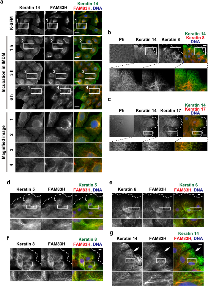
Figure 5. FAM83H is localized on keratin filaments extending to the cell-cell interface in HAM3 cells.
(a) In order to induce cell-cell attachments in HAM3 cells, culture medium was replaced from K-SFM medium to IMDM medium, and cells were then incubated in IMDM medium for 1, 3, or 6 h. Cells were stained with anti-keratin 14 and anti-FAM83H. (b–g) HAM3 cells were cultured in IMDM medium for 24 h and then stained using the indicated antibodies. The edge of cell sheets was indicated by dotted lines. In (g), an arrow indicates the cell-spreading region. In all images, DNA was stained by DAPI (blue), the magnified images at the regions enclosed by squares are shown in the margin, and scale bars indicate 20 μm (a–c) or 10 μm (d–g). Ph, phase-contrast.

