
Fig 2.
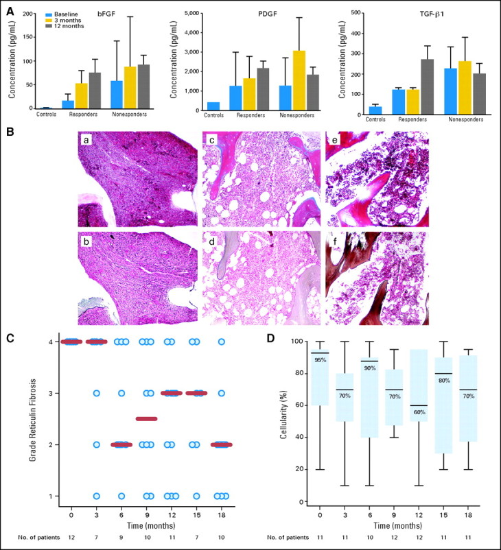
Effect of lenalidomide and prednisone therapy on cytokine levels, reticulin fibrosis and bone marrow cellularity in responders. (A) Levels of transforming growth factor beta (TGF-β), platelet-derived growth factor (PDGF), and basic fibroblast growth factor (bFGF) were measured by enzyme-linked immunosorbent assays in peripheral blood before study entry and after 3 and 12 months of therapy in nine responders and 21 nonresponders to lenalidomide and prednisone. Control levels represent mean values in peripheral blood obtained from eight healthy volunteers. (B) Effects of lenalidomide and prednisone on the bone marrow of a responder carrying the JAK2V617F mutation. The upper panels (a, c, e) represent collagen trichrome stains and the lower panels (b, d, f) depict reticulin silver stains at baseline, and after 6 and 12 months of therapy. At baseline, the bone marrrow is markedly hypercellular (100%) with minimal increase in (a) collagen but with a diffuse, dense increase in reticulin fibers with (d) extensive intersections corresponding to a fibrosis score of 4. After 12 months of therapy, the bone marrow cellularity decreased to 70% with only a focal residual network of reticulin fibers in (f) perivascular areas corresponding to a fibrosis score of 1. (C) The dynamics of reticulin fibrosis are shown. All 11 assessable responders had a pretreatment reticulin fibrosis score of 4. This score was reduced to at least 2 in 10 of them during therapy (P = .007). (D) Changes in bone marrow cellularity among responders with respect to pretreatment values were not significant (P = .80).