
Fig. 3.
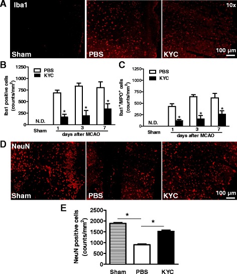
Effects of KYC on microglia activation and loss neuron in the brain of mice. MCAO mice were prepared as described in Fig. 2. a Images of Iba1+ in brain cortex ischemic core 3 days after MCAO; b Counts of Iba1+ cells in the brain sections of mice 1, 3, and 7 days after MCAO. c Counts of Iba1+/MPO+cells in the brain sections of mice 1, 3, and 7 days after MCAO. All data represented n = 4 (p < 0.05, KYC vs. PBS group, t test). d Images of immunostaining of neuron (NeuN) in brain cortex ischemic core 3 days after MCAO; e Counts of NeuN positive cells (n = 5/group, *p < 0.05, one-way ANOVA with Bonferroni test)