
FIGURE 3.
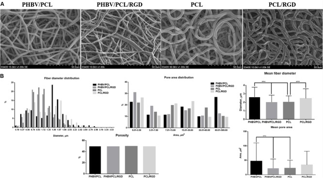
PHBV/PCL and PCL vascular grafts with RGD peptides have a distinct morphology. (A) Scanning electron microscopy images of PHBV/PCL and PCL grafts with and without RGD peptides. (B) Morphological parameters of PHBV/PCL and PCL grafts with and without RGD peptides; quantitative image analysis revealed a higher mean fiber diameter and mean pore area in PHBV/PCL compared to PCL grafts and in PHBV/PCL grafts with RGD peptides compared to those without, data are represented as mean with standard deviation, ∗∗∗P < 0.001, two-tailed Student’s t-test.