
FIGURE 5.
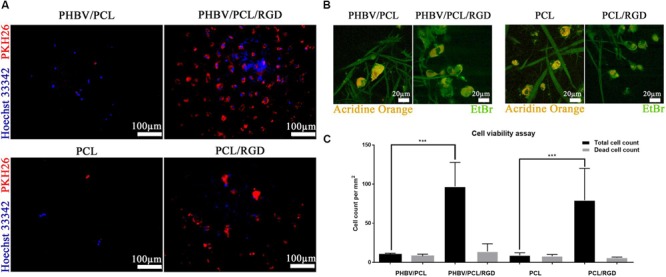
PHBV/PCL and PCL scaffolds with RGD peptides improve cell viability. (A) Fluorescence microscopy showed several-fold increase in total cell count on PHBV/PCL and PCL scaffolds with RGD peptides in comparison with those without. Nuclei and cytoplasm are stained blue and red, respectively. (B) Confocal laser scanning microscopy confirmed the results obtained by fluorescence microscopy. Nuclei of the dead cells are stained orange whilst viable cells are stained green. (C) Quantitative analysis confirmed the results of microscopy analysis, data are represented as median with interquartile range, ∗∗∗P < 0.001, Mann–Whitney U-test.