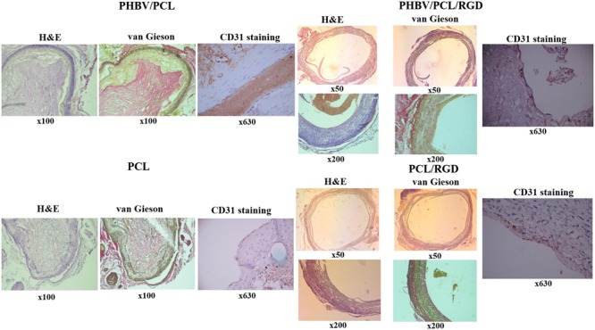
FIGURE 6.

Modification of PHBV/PCL and PCL vascular grafts with RGD peptides enhances formation of endothelial cell monolayer. H&E and van Gieson staining revealed a putative endothelial cell monolayer on PHBV/PCL and PCL grafts with RGD peptides but not on those without; this was further confirmed by CD31 staining, which identified a monolayer of CD31-positive cells (brown) at the inner surface of both polymer grafts with RGD peptides.
