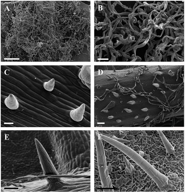
Figure 2.

SEM micrographs of ribbon and simple trichomes. (A,B) Ribbon trichomes on abaxial leaf surfaces of a young leaf in V. wilsoniae. (C–F) Simple trihcomes: (C) young simple trichomes on veins of V. romanetii; (D,E) simple trichomes on veins of young leaves in V. davidii; (F) simple trichomes on adaxial leaf surfaces of V. retordii. Scale bars: (A) = 500 μm; (B,E) = 50 μm; (C) = 20 μm; (D,F) =100 μm.
