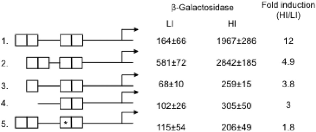
Figure 4.
Altering the IdeR binding region decreases iron dependent induction of PbfrB. The intact or modified DNA sequences upstream of bfrB containing the four iron boxes (IB1-4) were fused to lacZ and the fusions were individually introduced into Msm. β-galactosidase activity in cells grown in low and high iron medium as described in experimental procedures is shown as well as the fold induction in high iron relative to low iron. 1 to 5 indicates the modifications in the IdeR binding region: 1. The intact IdeR binding region. 2. A 5 bp deletion was made in the spacer sequence between IB2 and IB3. 3. IB1 was deleted. 4. IB1 and IB2 were deleted. 5. *IB3 was mutagenized (AGCCTT changed to CTATGC). The background activity of strains transformed with the vector alone was subtracted from the total activity. The values represent means ± SD from biological triplicates.

