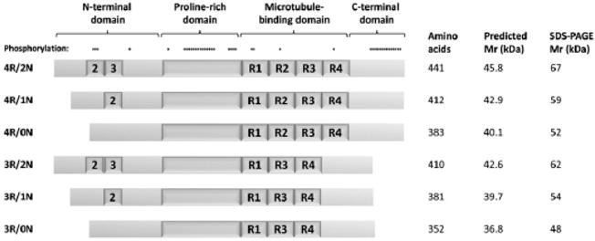
Fig.2.

Isoforms of the microtubule-associated protein Tau. The schematic depicts the six Tau isoforms expressed in the adult human brain, labeled to the left of each protein. Positions of major protein domains are shown above the longest isoform. The N-terminal insertions encoded by exons 2 and 3, and the four tandem repeats (R1–4) in the microtubule-binding domains are indicated. Sites of known phosphorylation are shown above the longest isoform The table to the right of the figure shows the number of amino acids, predicted molecular mass and electrophoretic mobility of each protein isoform.
