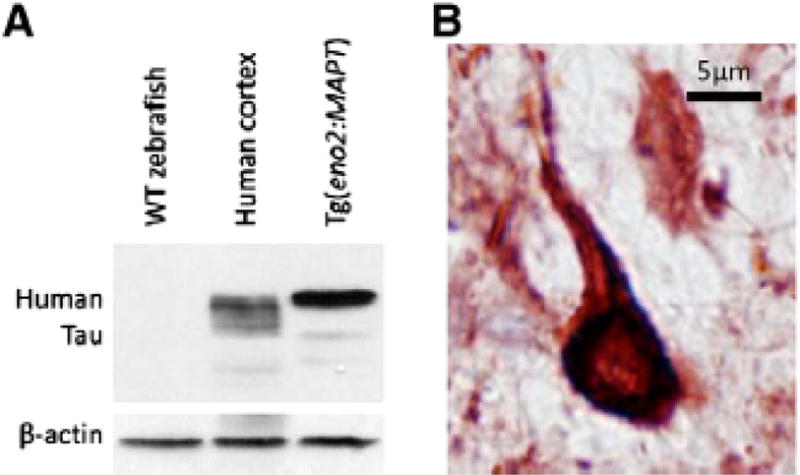
Fig. 5.

Human Tau expression in Tg(eno2:MAPT) zebrafish neurons. A: A western blot was made with protein lysate from wild-type zebrafish brain (lane 1), control post-mortem human cortex (lane 2) and Tg(eno2:MAPT) brain (lane 3). The blot was probed using an antibody to human Tau (upper panel) followed by an antibody to β-actin (lower panel). Abundant expression of human 4R-Tau is seen in transgenic zebrafish compared with the six isoforms in normal human brain [111]. B: Sections of Tg(eno2:MAPT) zebrafish brain were labeled using the antibody to human Tau used in the western blot experiment shown in panel A and a histochemical reaction yielding a red product. The micrograph shows a brainstem neuron with dense Tau immunoreactivity in the cell body and proximal axon, resembling neurofibrillary tangles shown in Fig. 1B [111].
