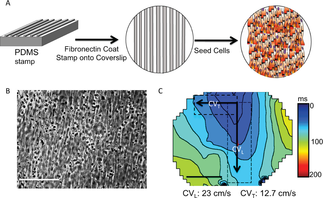
Figure 1. Patterned NRVM monolayers.
(A) NRVMs were plated onto cover slips coated with fibronectin lines (B). Light microscopy shows myocytes growing in a longitudinal direction (white bar=10µm). (C) Patterned NRVMs had anisotropic conduction, faster in the longitudinal (CVL) than the transverse direction (CVT). (n=3)

