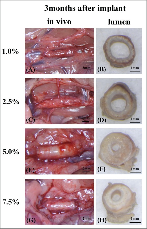
FIGURE 4.

The grafts 3 months after implant just before removed from rat abdominal aortic (A, C, E, G). It was able to confirm much tissue around grafts so that coating density was low. Grafts lumen at the mid-portion were checked after removed from rat (B, D, F, H). There were many coating around grafts in high density. Also lumen became thick than low density grafts because of neointimal hyperplasia (F, H). On the other hand in low density grafts, there were no neointimal hyperplasia and thin inner layer were confirmed and most of coating were decomposed (B, D).
