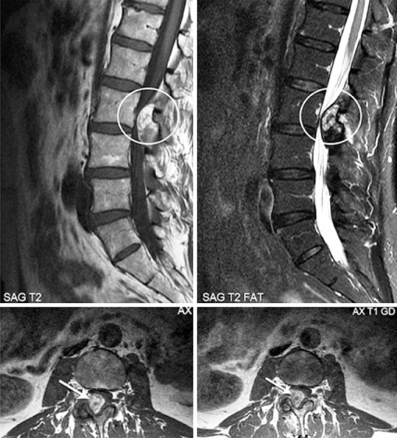
Figure 1. Magnetic resonance imaging showing heterogeneous intraspinal and extradural mass, contiguous to the interapophyseal joint at L2-L3, on the right, with radicular and dural compression, with no signs of fat. Parietal enhancement after contrast.

