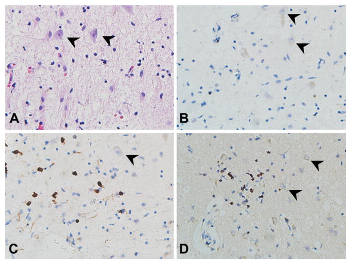
Fig. 3.
Sections from the hippocampus/entorhinal cortex showing T-lymphocytic inflammation around neurons (arrowheads, 40×). A. Hematoxylin and eosin staining. B. CD4 immunopositivity was present in only a few cells. C. CD8 and D. TIA-1 (a granule-associated protein of cytotoxic T-cells and NK cells) immunostaining was prevalent among the T-lymphocytes, supporting a cytotoxic subtype.

