
Figure 7-7.
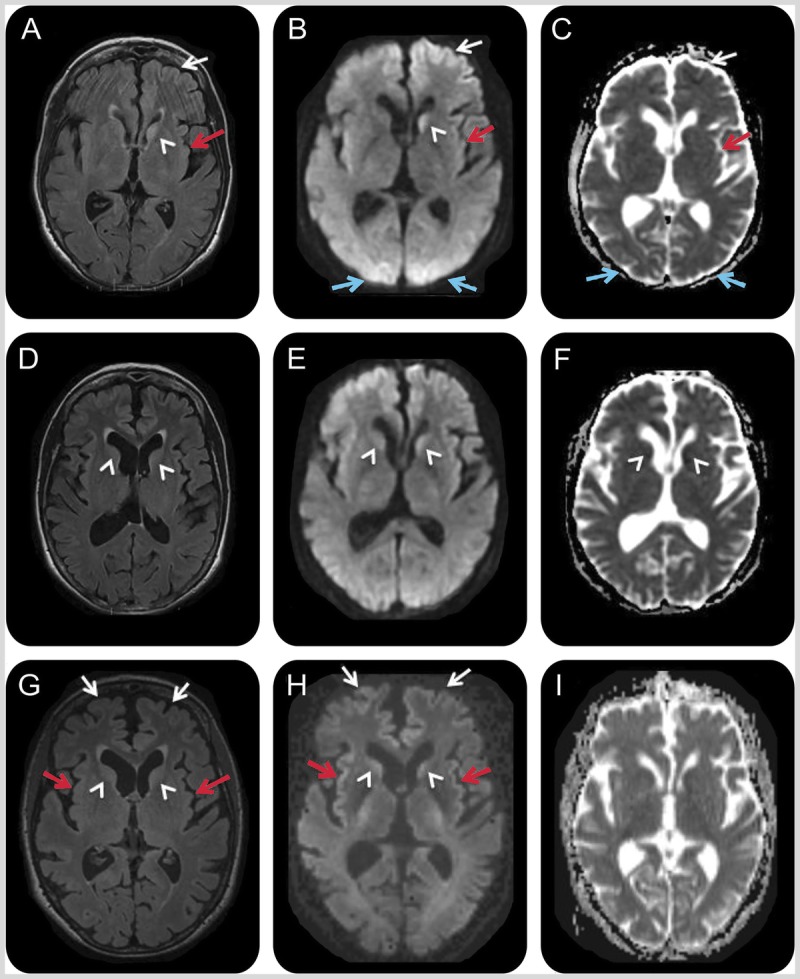
MRI of the patient in Case 7-2. A 66-year-old woman with hypoglycemic encephalopathy. Fluid-attenuated inversion recovery (FLAIR) (A, D, G), diffusion-weighted images (DWI) (B, E, H), and apparent diffusion coefficient (ADC) map (C, F, I) sequences 2 days (A–C), 3 weeks (D–F), and 1 month (G–I) after onset. Initial MRI (A–C) showed left frontal (white arrows), left insular (red arrows), bilateral medial occipital (blue arrows), and left caudate (white arrowhead) FLAIR/DWI hyperintensity with restricted diffusion, which is subtle but definitely appreciable. Repeat MRI about 3 weeks later (D–F) showed possible reduced FLAIR/DWI hyperintensity in the left caudate head and medial occipital regions, and possible increased right caudate FLAIR hyperintensity and restricted diffusion (D–F; white arrowheads). A third MRI 1 week later, 1 month after onset (G–I), revealed more intense FLAIR/DWI insular (G, H; red arrows) and frontal cortical hyperintensities (G, H; white arrows) and possible restricted diffusion and FLAIR hyperintensity still present in the caudate heads (G, H; arrowheads). The resolution of occipital cortical ribboning in such a short time argued against a diagnosis of sporadic Jakob-Creutzfeldt disease.
Reprinted with permission from Rosenbloom MH, et al, Neurol Clin Pract.50 cp.neurology.org/content/5/2/108.full. © 2015 American Academy of Neurology.