
Figure 7-8.
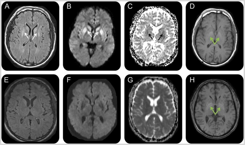
MRI of the patient in Case 7-3. A 50-year-old man with extrapontine myelinolysis. Initial MRI 2 months after onset (A–D) showed symmetric bilateral striatal fluid-attenuated inversion recovery (FLAIR) (A)/diffusion-weighted imaging (DWI) (B) hyperintensities (A, B; white arrows) with corresponding hypointensities on the apparent diffusion coefficient (ADC) map suggesting restricted diffusion (C; black arrows). Bilateral globus pallidus hyperintensities were present on T1-weighted images (D; green arrows). MRI 1 month later, 3 months after onset (E–H), showed resolution of the prior FLAIR (E), DWI (F), and ADC (G) map abnormalities but no change in the globus pallidus T1 hyperintensities (H; green arrows).
Reprinted with permission from Rosenbloom MH, et al, Neurol Clin Pract.50 cp.neurology.org/content/5/2/108.full. © 2015 American Academy of Neurology.