
Fig. 7.
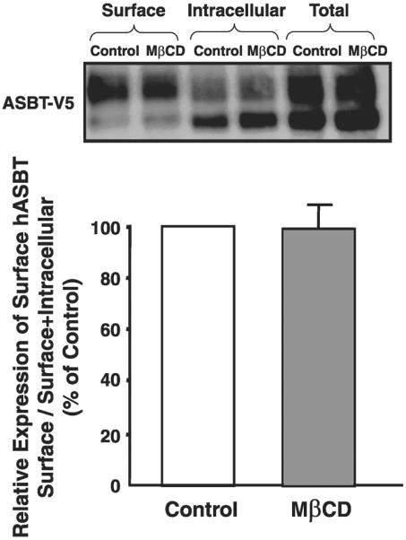
Plasma membrane expression of ASBT-V5 fusion protein in Caco-2 cells. Caco-2 cells were transfected with ASBT-V5 by electroporation utilizing Amaxa Nucleofector System as described in materials and methods; 24 h posttransfection, monolayers grown on plastic support were treated with 10 mM MβCD or vehicle alone for 1 h at 37°C and subjected to biotinylation at 4°C using sulfo-NHS-SS-biotin. Cells were then lysed, and surface biotinylated proteins were precipitated with streptavidin-agarose from equal amounts of total cellular protein. Precipitated proteins (surface) were separated on SDS-polyacrylamide gel electrophoresis and electroblotted to nitrocellulose blots. Western blotting analysis was performed with anti-V5. The relative abundance of ASBT-V5 fusion protein in the biotinylated fractions is shown and is expressed as the density of ASBT-V5 band normalized to the density of total hASBT-V5 (surface+intracellular). Data were obtained from 3 separate experiments.