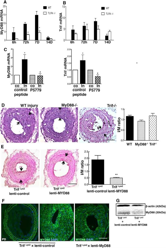
Figure 3.
Both myeloid differentiation primary response gene 88 (MyD88) and Trif contribute to intimal hyperplasia formation after wire injury. A–C, Arterial mRNA levels for MyD88 and Trif were measured by reverse transcription polymerase chain reaction and normalized to 18S transcript levels at indicated time points after wire injury. Relative mRNA levels compared with the uninjured carotid artery are displayed (n=4–6). D, Carotid arteries from wild-type (WT) mice (n=10), MyD88−/− mice (n=16), and Trif Lps2 mice (n=7) were analyzed at day 28 after arterial injury. E, Arterial intimal hyperplasia after local MyD88 silencing in TrifLps2 mice (Trif Lps2 +lenti-control, n=8; Trif Lps2+lenti-shMyD88, n=7) is shown. Intima and media area (I/M) was reduced in the lenti-shMyD88 group compared with the lenti-control. F, Focal MyD88 expression is shown in carotid arteries locally treated with lenti-control or lenti-shMyD88. Representative hematoxylin & eosin–stained and fluorescent-stained sections are shown. The I/M ratios were quantified by planimetry at 28 days. G, Tissue lysates from aortic arteries (30 μg) were prepared from lenti-shMyD88 and lenti-control at 28 days after application of lentivirus and subjected to Western blot with anti–high-mobility group box 1 and β-actin antibodies. Data represent the mean±SEM. *P<0.05, **P<0.01 vs controls, respectively. Scale bar, 100 μm.

