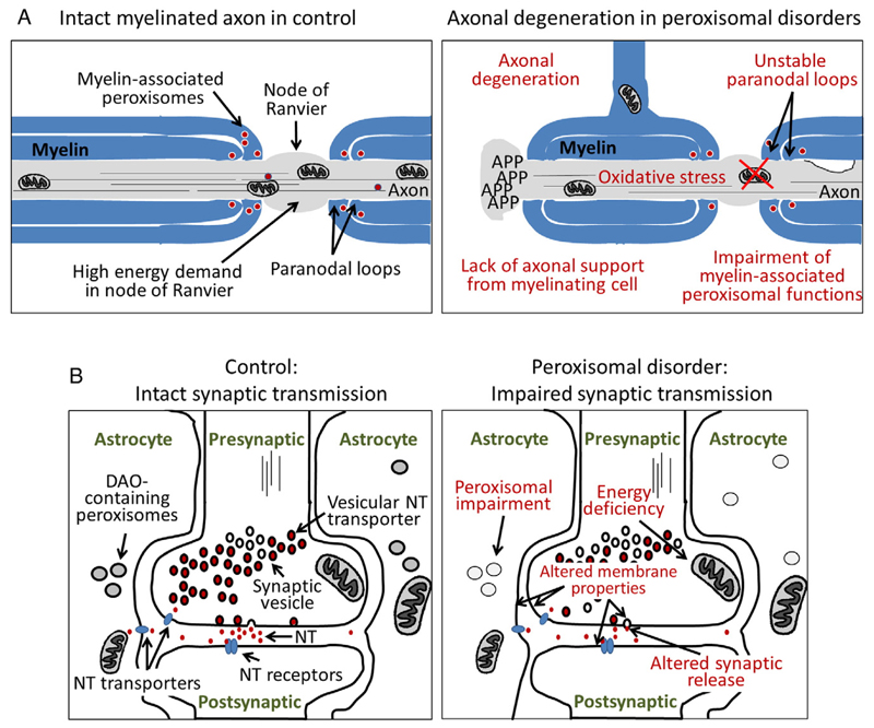
Fig.3.
Schematic representation of abnormalities of myelinated axons and synaptic transmission in peroxisomal deficiencies. (A) The left panel shows a myelinated axon at the level of a node of Ranvier in a healthy control. The myelin sheath of oligodendrocytes (in the CNS) or Schwann cells (in the PNS) surrounds and isolates the axon, except at the node of Ranvier allowing depolarization of the neuronal membrane and propagation of electrical signals. Note that a multitude of ion channels and Na+/K+-ATPases (not indicated) are located at the node of Ranvier and entail a high energy demand. In the right panel, different pathological features are indicated that may contribute to the axonal degeneration frequently observed in peroxisomal disorders, for example, adrenomyeloneuropathy (the late-onset variant of X-ALD). A scenario can be envisaged, where peroxisomal dysfunction and abnormal accumulation of lipid metabolites in myelinating cells lead to unstable paranodal loops and a loss of axonal support resulting in energy deficits and oxidative damage in the axons and progressive axonal degeneration. (B) A normal synapse with the surrounding astrocytes is depicted (left panel), representative for a synapse of any neurotransmitter. D-Amino acid oxidase is indicated for its role in D-serine degradation at e.g. glutamatergic synapses. The right panel shows several possible disturbances of synaptic function (red text) that could lead to altered neurotransmission, as predominantly described in ether lipid deficiency. NT, neurotransmitter; DAO, D-amino acid oxidase

