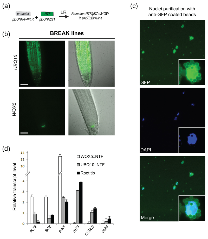
Figure 4. Nuclei purification using anti-GFP beads and a modified INTACT protocol.
(a) Cloning strategy to generate the BREAK lines.
(b) BREAK lines expression pattern for UBQ10∷NTF; WOX5∷NTF. Scale bars: 50 μm.
(c) Epifluorescence micrographs of affinity-captured nuclei. Note the autofluorescence of magnetic beads in the GFP channel. A higher magnification of a bead-bound nucleus is shown as an insert.
(d) RT-qPCR analyses of selected genes in INTACT-purified nuclei as well as root tips. Normalized transcript levels are given relative to AT5G60390 expression level arbitrarily set to 100. Error bars correspond to standard deviations from two biological replicates.

