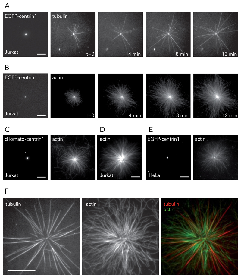
Figure 1. Cytoskeleton filament assembly from isolated centrosomes.
(A and B) Centrosomes were isolated from T lymphocytes expressing EGFP-centrin1 and seeded on glass coverslips. (A) The addition of purified tubulin dimers led to the assembly of dynamic microtubules. Time is in minutes. Images are representative of 7 independent experiments. (B) The addition of purified actin monomers led to the assembly of radial arrays of actin filaments. Time is in minutes. Images are representative of 12 independent experiments. (C, D and E) Centrosomes were isolated from Jurkat cells expressing dTomato-centrin1 (5 independent experiments) (C), non-modified Jurkat cells (5 independent experiments) (D) and from HeLa cells expressing EGFP-centrin1 (8 independent experiments) (E). All centrosome preparations induced the growth of actin filaments in the presence actin monomers. (F) The assembly of both microtubules and actin filaments from isolated centrosomes. Images are representative of 5 independent experiments. Scale bars:10 μm.

