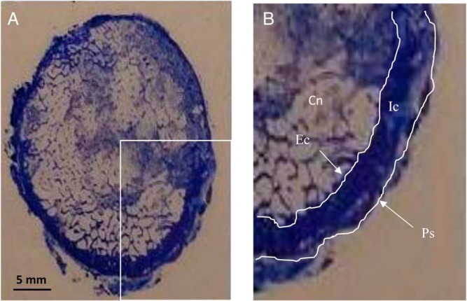
Figure 1.
A, Cross section of the femoral neck, toluidine blue stain. B, Magnified view of lower right quadrant of the FN indicating the four envelopes that were analyzed: Cn, cancellous; Ec, endocortical; Ic, intracortical; Ps, periosteal. The histomorphometric analysis was performed within each envelope for the entire cross section.

