
Figure 3.
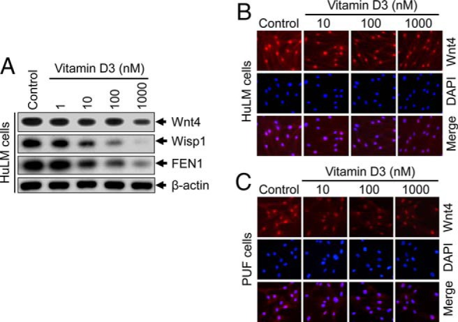
Effect of vitamin D3 on protein expression of Wnt4, Wisp1, and FEN1 in cultured human uterine fibroid cells. A, HuLM cells were serum starved and treated with increasing concentrations of vitamin D3 (0, 1, 10, 100, and 1000 nM) for 48 hours. Equal amounts of each cell lysate were analyzed by Western blots using anti-Wnt4, anti-Wisp1, and anti-FEN1 antibodies. β-Actin Western blot was used as loading control. B and C, Immunofluorescence analyses were performed using both HuLM cells (B) and human PUF cells (C) cultured on glass coverslips and treated with increasing concentrations of vitamin D3 (0, 10, 100, and 1000 nM) for 48 hours. Cells were fixed, permeabilized, and stained with anti-Wnt4 antibody (1:50 dilution) followed by incubating with carbocyanine 3-conjugated antirabbit secondary antibody. Wnt4 staining (red) was monitored by fluorescence microscopy. Nuclei of cells were stained with 4′, 6-diamino-2-phenylindole. Pictures were taken at ×200 magnification.