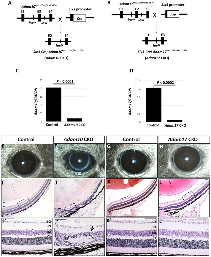
Fig 2. Conditional ablation of Adam10 and Adam17 in the retina.
(A) Mice carrying homozygous floxed Adam10 allele (Adam10tm1.1Zhu) were crossed with Six3-Cre transgenic mice expressing Cre in the optic cup and optic stalk resulting in conditionally ablated Adam10 in the retina. (B) A similar approach was taken to generate Adam17 CKO mice after homozygote Adam17tm1.2Bbl mice were crossed to Six3-Cre mice. (C) Semi-quantitative analysis of the relative expression of Adam10 transcript containing exon 3 normalized to Gapdh expression from RNA isolated from newborn control and Adam10 CKO littermates revealed significantly reduced (P<0.0001; n = 3) levels in Adam10 CKO retinae when compared to the controls. (D) Semi-quantitative analysis of the relative expression of exon 2 containing Adam17 mRNA normalized to Gapdh expression from RNA isolated from newborn control and Adam17 CKO retinae also revealed significantly reduced (P<0.0002; n = 3) levels in Adam17 CKO retinae when compared to the controls. In both (C) and (D), the bars represent mean values ± SEM. Significance was established following Student’s t-test analysis and P<0.05 was considered significant. (F) Adam10 CKO eyes appeared smaller when compared to the eyes in the control mice (E) whereas Adam17 CKO eyes (H) that did not appear to differ from age-matched control littermates (G). H&E staining of Adam10 CKO eyes at 8 weeks of age (J) revealed severe retinal abnormalities characterized by the formation of rosettes (J’, arrow) in contrast to the retinal laminae in the age-matched controls (I-I’). Histological analysis of Adam17 CKO eyes (L-L’) did not identify any morphological differences when compared to the age-matched controls (K-K’). Figures shown in (I’) through (L’) are enlarged images of the areas depicted by the dashed boxes in Figures (I) through (L). GCL = ganglion cell layer, IPL = inner plexiform layer, INL = inner nuclear layer, OPL = outer plexiform layer, ONL = outer nuclear layer. Scale bars = 50 μm.

