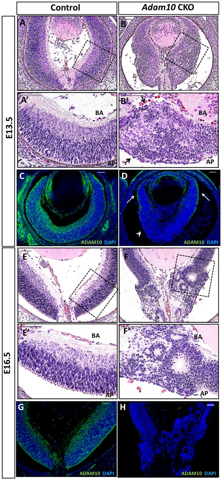
Fig 3. Retinal morphology in control and Adam10 CKO mice.

H&E staining revealed first abnormalities in E13.5 Adam10 CKO retinae (B) within the central region characterized by rounded and highly disorganized nuclei along the apical-basal axis resulting in disrupted basal and apical surfaces (B’, arrows) in contrast to the highly organized control retinae (A-A’). ADAM10 immunostaining of E13.5 Adam10 CKO retinae (D, arrowhead) revealed an absence of ADAM10 expressions in most cells within the central retina when compared to the age-matched controls (C). However, residual ADAM10 expression was detected in the peripheral retinae of Adam10 CKO mice (D, arrows). H&E staining of E16.5 Adam10 CKO retinae (F) identified within the central region rosettes of various sizes (F’) in contrast to the highly organized retinal laminae in the age-matched control retinae (E-E’). Immunostaining of E16.5 Adam10 CKO retinae (H) did not identify any ADAM10-positive cells in contrast to the age-matched controls (G). BA = basal surface, AP = apical surface. Scale bars = 50 μm.
