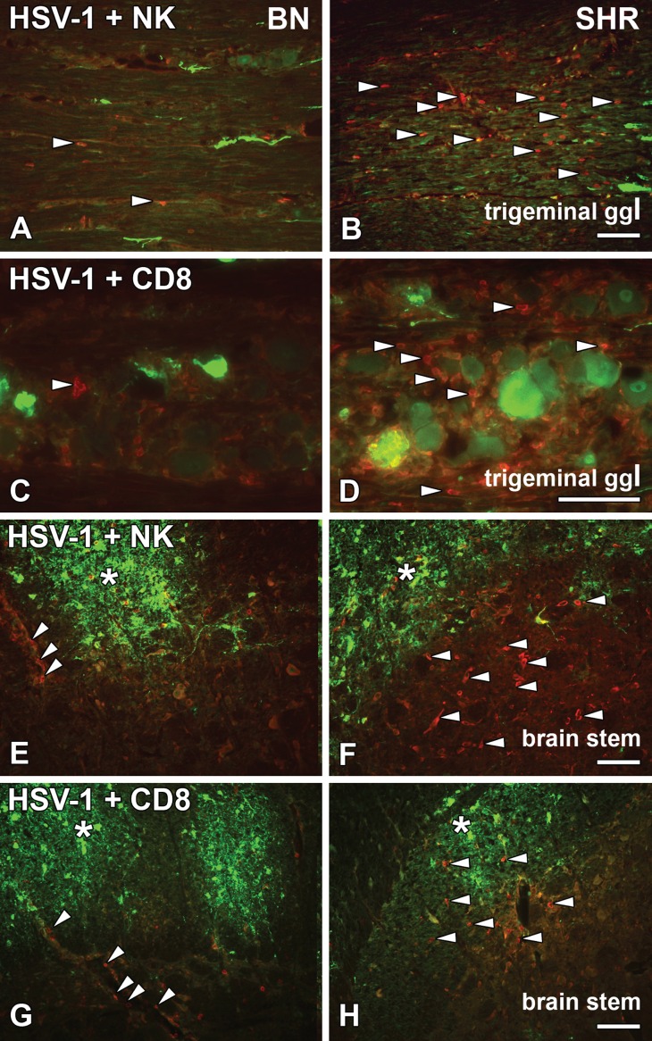
Fig 2. HSV-1 activated NKR+ and CD8+ cells in the trigeminal ganglia and brain stem.
Longitudinal/sagittal sections of the trigeminal ganglia (A–D) and transversal/coronal sections from the brain stem (E–H) from HSV-1 infected BN and SHR rats at 4dpi, stained for HSV-1 marker (green) (A–H), NK cell marker NKR (red) (A, B, E and F) and cytotoxic T cell marker CD8 (red) (C, D, G and H). In the trigeminal ganglia, NK cells were less visible among the axons infected with HSV-1 in the resistant BN rats (A; arrowheads) compared to the SHR rats (B; arrowheads). Also, fewer CD8+ cells were observed in BN (C; arrowhead) rats as compared to the SHR (D; arrowheads). While in the brainstem, HSV-1 staining could be seen in both BN (E and G; asterisk) and SHR (F and H; asterisk). NKR+ cells were less visible in BN rats (E; arrowheads) outlining the blood vessels as well as cytotoxic CD8+ cells (G; arrowheads). In SHR rats, more NK cells (F; arrowheads) and CD8+ (H; arrowheads) were present in the parenchyma. Scale bar: 50 μm = (A = B = E = F = G = H), (C = D).

