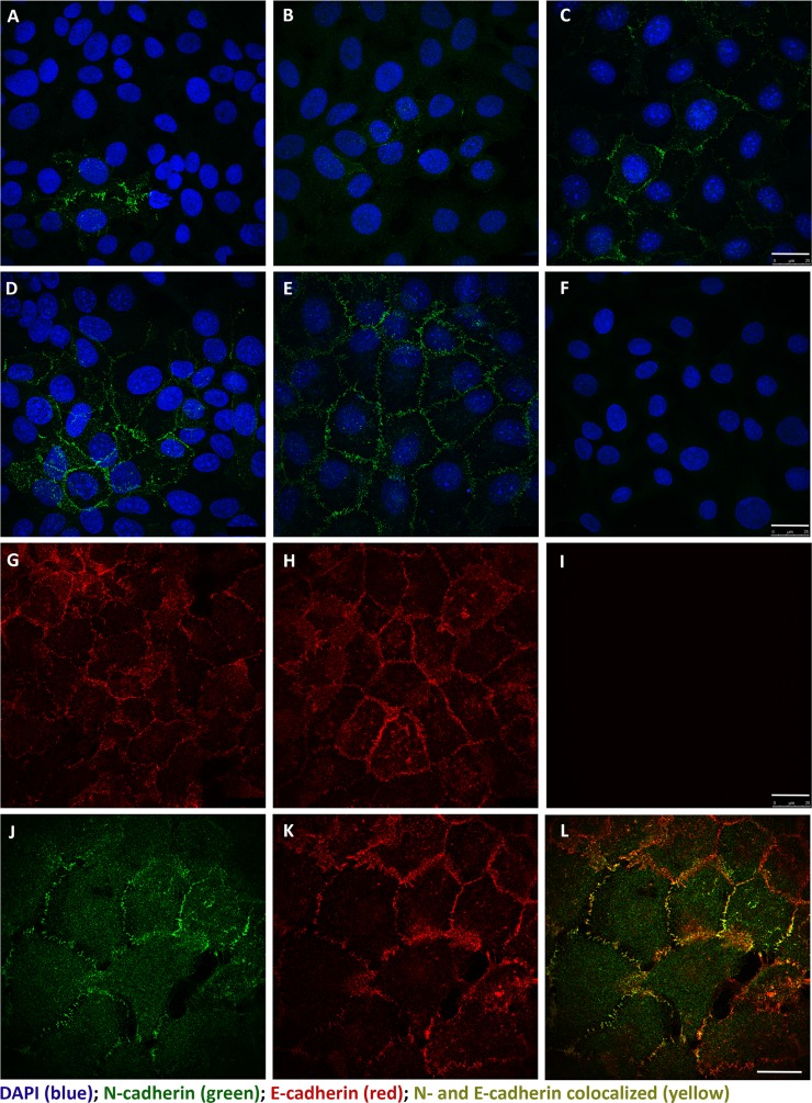
Fig 6. Immunofluorescent localization of N- and E-cadherin in UROtsa cells by scanning confocal microscopy.
N-cadherin (green) was found to be localized to the plasma membrane, when present, in the parent and transformed UROtsa cell lines: (A) UROtsa parent; (B) Cd#1; (C) Cd#5; (D) As#3; (E) As#6 with DAPI shown as a counterstain. A representative negative control that lacks primary antibody is shown in (F) with the UROtsa Parent cell line. E-cadherin (red) was also found to be localized to the plasma membrane and present in nearly all of the cells within the population. Representative images are shown for UROtsa Parent (G) and As#6 (H) cell lines. Likewise, a representative negative control that lacks primary antibody is shown in (I) for the UROtsa parent cells. To determine if N- and E-cadherin co-localized within the same cells, a single 0.347 μm z-plane image is shown for As#6 (J-L). N-cadherin (green) is shown in (J), E-cadherin (red) is shown in (K), and the merged images showing co-location of N- and E-cadherin (yellow) is shown in (L). Both scale bars = 25 μm and are shown in (I) for panels A-I and in (L) for panels J-L.

