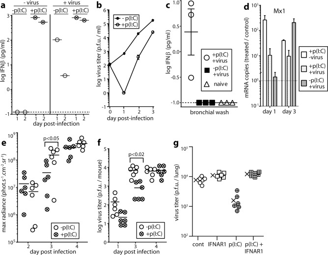
Fig 1. IFNαβ induction by and effect of IFNαβ induction on MuHV-4 infection in vitro and in vivo.
a. RAW-264 monocyte/macrophages were infected or not with MuHV-4 (2 p.f.u. / cell) then cultured ± poly(I:C) as a positive control of IFN-I induction (50μg/ml). Cell supernatants pooled from triplicate cultures were assayed for IFNβ by ELISA 1 and 2d later. The dashed line shows the limit of assay sensitivity. A replicate experiment gave equivalent results. b. RAW264 cells were infected and cultured ± poly(I:C) to induce IFN-I as in a. Supernatants were assayed for infectious virus by plaque assay. Symbols show mean ± SEM of triplicate cultures. IFN-I induction significantly reduced virus titers from d1 onwards (p<0.03). c. Mice were given i.n. MuHV-4 (3x10⁴ p.f.u.), with or without poly(I:C) inoculation (50μg i.p. and i.n., 6h before and at the time of virus inoculation) as a positive control of IFN-I induction. After 1d bronchial washes were assayed for IFNβ by ELISA. Each point shows 1 mouse. Bars show mean ± SEM. The dashed line shows the limit of assay sensitivity. Poly(I:C) significantly increased IFNβ production in infected mice (p<0.01). d. Mice were infected i.n. or not with MuHV-4 with or without poly(I:C) treatment as in c. 1 and 3d later Mx1 mRNA in lungs was quantitated by real time PCR and normalized by cellular nidogen-1 copy number. Numbers show the fold increase in Mx1 copy number of treated mice over the untreated controls (dashed horizontal line). Bars show mean ± SEM of 3 mice. Mx1 copies were significantly increased by poly-IC at d1 (p<0.05) but not at d3. e. Mice were infected i.n. with MHV-LUC, and treated or not with poly(I:C) as in c. Lung infection was tracked by luciferin injection and live imaging of light emission. Circles show individual mice, bars show means. Poly(I:C) significantly reduced luciferase counts at d3 but not at d2 or d4. f. Mice were infected i.n. with MuHV-4 and treated or not with poly(I:C) to induce IFN-I as in c. Lungs were titered for infectious virus by plaque assay. Circles show individual mice, bars show means. Poly(I:C) significantly reduced infection at d3 but not at other time points. g. Mice were given an anti-IFNAR blocking antibody or not (200μg i.p.) 24h before and poly(I:C) or not (50μg i.p. and i.n.) 6h before i.n. infection with MuHV-4 (3x10⁴ p.f.u.). 3d later lungs were titered for infectious virus by plaque assay. Cont = virus only. Crosses show means, other symbols show individual mice. Poly(I:C) significantly reduced titers (p<0.01) and this effect was reversed by anti-IFNAR antibody.

