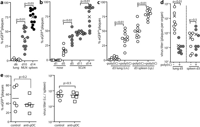
Fig 2. MuHV-4 propagation through IFNαβ-responding cells.
a. Mx1-cre mice were given MHV-4-RG i.n. (3x10⁴ p.f.u. in 30μl under anesthesia). Viruses were recovered from lungs by plaque assay and from lymphoid tissue by intact cell explant onto BHK-21 cell monolayers. Plaques were then typed as eGFP+ (switched) or mCherry+ (unswitched). For each mouse (circles), % eGFP+ = % switched of total recovered plaques. Crosses show means. MLN = mediastinal lymph nodes. b. Mice were given MHV-RG i.n. (3x10⁴ p.f.u. in 5μl without anesthesia) to infect just the nose, then analysed for viral fluorochrome switching as in a. SCLN = superficial cervical lymph nodes. Viruses were recovered from noses by plaque assay and from SCLN by intact cell explant (infectious centre assay). c. Mice were given i.n. or i.p. MHV-RG, with or without poly(I:C) (50μg i.n. or i.p. 6h before and at the time of infection) to maximally induce IFN-I. Viruses recovered 3d later from lungs by plaque assay (i.n.) or from spleens by infectious centre assay (i.p.) were analysed for fluorochrome expression as in a. d. Mice were infected i.n. (lung) or i.p. (spleen) and given poly(I:C) or not as in c. Virus titers were determined 3d later by plaque assay. Circles show individuals, crosses show means. e. Mx1-cre mice were depleted or not of pDCs with mAb 120G8, then infected i.p. with MuHV-RG (105 p.f.u.). 5d later spleens were titered for total recoverable virus by explant of intact splenocytes onto BHK-21 cells. Infectious centres (ICs) were also typed as mCherry+ (unswitched) or GFP+ (switched). % eGFP+ = % of total plaques that were switched. Bars show mean ± SEM, other symbols show individual mice. pDC depletion had no significant effect.

