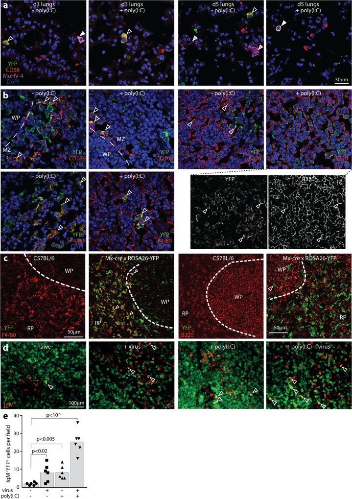
Fig 4. IFNαβ responses measured by Mx1-cre activation of ROSA26-YFP.
a. Mx1-cre x ROSA26-YFP mice were infected i.n. with MuHV-4 (3x10⁴ p.f.u.). IFN-I was induced or not with poly(I:C) (50μg i.n. and i.p., 6h before and at the time of infection). Lungs were harvested at d3 and d5 and stained for YFP, CD68 and MuHV-4 antigens. Nuclei were stained with DAPI. White arrows show example YFP+CD68+MuHV-4+ AMs; open arrows show YFP+CD68+ AMs; Grey-filled arrows show YFP-MuHV-4+ AEC1s. No AEC1s were YFP+. All YFP+ cells were AMs (n>30). Images are representative of 5 sections from each of 3 mice per group. b. Mx1-cre x ROSA26-YFP mice were infected i.p. with MuHV-4 (105 p.f.u.). IFN-I was induced or not with poly(I:C) (50μg i.p., 6h before and at the time of infection). D5 spleen sections were stained for YFP and CD169 (MZ macrophages), F4/80 (RP macrophages) or B220 (B cells). Nuclei were stained with DAPI and the cells visualized by confocal microscopy. Arrows show example YFP+ cells expressing the relevant cellular marker. For CD169 staining a dashed line shows the boundary between the B cell-dominated WP and the macrophage-dominated MZ, with CD169+ MZ macrophages lying adjacent to WP B cells. For B220 staining after virus + poly(I:C), separate channels are shown to make clear the extensive YFP expression in B cells. Images are representative of 5 sections from each of 3 mice per group. >50% of each macrophage population was YFP+, with no difference between poly-IC treated and untreated. In WP follicles, 10–50% of B220+ B cells were YFP+ after poly(I:C) treatment and 5–20% were YFP+ without poly(I:C) treatment. Quantitation for IgM+ B cells is shown in e. c. Naïve C57BL/6 and Mx1-cre x ROSA26-YFP spleen sections were stained for YFP, B220 and F4/80. YFP expression was evident in >80% of F4/80+ macrophages and <5% of B220+ B cells. Dashed lines show the MZ demarcation between F4/80+ RP macrophages and B220+ WP B cells. d. Mx1-cre x ROSA26-YFP mice were infected as in b. Spleen sections were stained for YFP and IgM to identify MZ B cells, and visualized by epifluorescence microscopy. Arrows show example YFP+IgM+ cells. e. Quantitation of YFP+IgM+ spleen cell numbers across 3 sections from each of 2 mice per group (5 fields of view per section). YFP expression in IgM+ B cells was significantly induced by both infection and poly(I:C). Bars show means, other symbols show individual mice.

