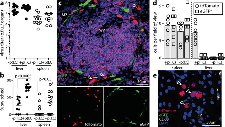
Fig 7. Mx1-cre-dependent fluorochrome switching of MCMV.
a. Mx1-cre mice were infected i.p. with MCMV-GR, which switches fluorochrome expression when exposed to cre recombinase. IFN-I was induced or not by i.p. poly(I:C) inoculation (pIC, 50μg / mouse) 6h before and at the time of infection. 5d later virus in livers and spleens was titered by plaque assay. Circles show individuals, bars show means. Poly(I:C) had no significant effect on titers (p>0.5). b. The samples from a were assayed for viral fluorochrome switching by identifying plaques as eGFP+ or tdTomato+. c. Example image from a MCMV-GR-infected and poly(I:C)-treated spleen shows eGFP+ and tdTomato+ cells around a WP follicle. Quantitation is shown in d. d. Quantitation of eGFP+ and tdTomato+ cells on liver and spleen sections, pooled from 3 mice per group infected as in a. Bars show means, other symbols show individual sections. Poly(I:C) significantly increased infected cell fluorochrome switching in livers (p<0.01) but not in spleens (p>0.5). e. Example image of a MCMV-GR-infected, poly(I:C)-induced, liver showing only tdTomato+ cells that do not co-localize with CD68. Two other livers gave equivalent results.

