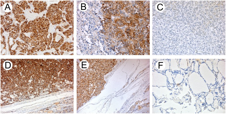
Fig 4. LAT1 immunohistochemistry.
(A-B) Pheochromocytoma, strong cytoplasmic staining of LAT1 with membranous enhancement sparing of the neoplastic nuclei and normal adrenal tissue (C). (D-E) Medullary thyroid carcinoma, strong cytoplasmic staining of LAT1 with membranous enhancement sparing of the neoplastic nuclei and normal thyroid tissue (F). (A-C and F) 20× original magnification; (D-E) 10× original magnification.

